Shoulder Muscles Diagram Anterior / Ever-Green Massage Therapy: Muscles of the Shoulder and Arm / Learn their origins/insertions, functions & exercises.
Shoulder Muscles Diagram Anterior / Ever-Green Massage Therapy: Muscles of the Shoulder and Arm / Learn their origins/insertions, functions & exercises.. Let's start by the anterior view of the diagram. The shoulder girdle consists of the clavicle (collar bone) and the scapula (shoulder blade) which generally move together as a unit. In this image, you will find muscles of the shoulder and chest, trapezius muscle, deltopectoral triangle, acromion you will also find deltoid muscle, cephalic vein, long head of biceps brachii muscle, short head of biceps brachii muscle, triceps brachii muscle, latissimus dorsi muscle, serratus anterior. Shoulder diagram to mainly explain you about how your shoulder work and to describe every inner part of your shoulder including muscles, joints, and every human has a body part called shoulder. Anterior graphic of the shoulder.
Cuff muscles of the back shoulder muscles and chest human anatomy diagram the shoulder muscles bridge the transitions from the torso into the head neck area and into the upper extremities of the arms and hands shoulder muscle names human anatomy diagram structuring bones anatomy. Anterior superior iliac spine insertion: Deltoid (posterior fibers), teres major, teres minor, latissimus dorsi, pectoralis major (sternocostal fibers). Superficial layer with deltoid, trapezius, pectoralis. There are around 650 skeletal muscles within the typical human body.
Anterior Torso Muscle Anatomy from cdn.thinglink.me • exion of the shoulder • adduction of the shoulder • horizontal adduction of the shoulder. Diagram shoulder muscles anatomy 101 shoulder muscles the handcare blog. Learn faster with interactive shoulder quizzes, diagrams and worksheets. The shoulder joint (glenohumeral joint) is a ball and socket joint between the scapula and the the resting tone of these muscles act to compress the humeral head into the glenoid cavity. They're all small, simple muscles. Rotator cuff is formed by a group of four muscles that surround the shoulder joint. Start studying shoulder muscles (anterior). Superficial layer with deltoid, trapezius, pectoralis.
The muscles of the anterior shoulder girdle include in fact, this muscle can actually be thought of three individual muscle compartments consisting of an anterior portion, a middle portion, and a posterior portion.
Now label the diagram in your workbook! Superficial layer with deltoid, trapezius, pectoralis. These muscles form the outer shape of the shoulder and underarm. Only the clavicle connects directly to the rest of the. Printable shoulder muscles diagrams to help you study the muscles structure in human's shoulder. Anterior border of lateral 1/3rd of clavicle. Medial tibia near tibial tuberosity action: Shoulder stretching exercises, including anterior shoulder stretch, chest stretch, triceps… Learn vocabulary, terms and more with flashcards, games and other study tools. The anterior, lateral and posterior deltoid heads. Diagram shoulder muscles anatomy 101 shoulder muscles the handcare blog. Start studying shoulder muscles (anterior). Muscles of the shoulder can be subdivided into a variety of groups depending on origin, topography, function or innervation.
The muscles labelled in the anterior muscles diagram shown above are listed in bold in the following table sternocleidomastoid trapezius serratus anterior latissimus dorsi pectoralis major pectoralis minor (deep muscle) rectus abdominus external oblique internal oblique transversus abdominus. Anterior superior iliac spine insertion: Mri, radiographical, and illustrated anatomical. The thickened middle ghl should not be confused with. Let's start by the anterior view of the diagram.
Shoulder & Arm - Atlas of Anatomy from doctorlib.info Medical illustration of the shoulder's muscles : Although three ligaments protect and surround the shoulder joint, most of its stability comes from the powerful muscles and tendons of the rotator cuff. We have five muscle diagrams of the shoulder. Only the clavicle connects directly to the rest of the. The shoulder has about eight muscles that attach to the scapula, humerus, and clavicle. Posterior part of the deltoid: • coracobrachialis • pectoralis major • subscapularis. This is a table of skeletal muscles of the human anatomy.
Deltoid (anterior fibers), pectoralis major (clavicular fibers), coracobrachialis, biceps.
The shoulder joint is supplied by the anterior and posterior circumflex humeral arteries, which are both. Shoulder diagram to mainly explain you about how your shoulder work and to describe every inner part of your shoulder including muscles, joints, and every human has a body part called shoulder. The system used here groups the muscles based on their function and topography (which are closely related in the upper limb) If deltoid is paralysed, rounded contour of the shoulder is lost and there is loss of power of abduction of arm from 15 to 90ο. The shoulder joint has the most range of motion of any joint on the. Right anterior basal segmental bronchus. The anterior, lateral and posterior deltoid heads. There are around 650 skeletal muscles within the typical human body. The thickened middle ghl should not be confused with. Flexes and medially rotates arm; Let's start by the anterior view of the diagram. Muscles of the shoulder can be subdivided into a variety of groups depending on origin, topography, function or innervation. Learn vocabulary, terms and more with flashcards, games and other study tools.
Rotator cuff is formed by a group of four muscles that surround the shoulder joint. The muscles of the anterior shoulder girdle include in fact, this muscle can actually be thought of three individual muscle compartments consisting of an anterior portion, a middle portion, and a posterior portion. Mri, radiographical, and illustrated anatomical. Printable shoulder muscles diagrams to help you study the muscles structure in human's shoulder. Medial tibia near tibial tuberosity action:
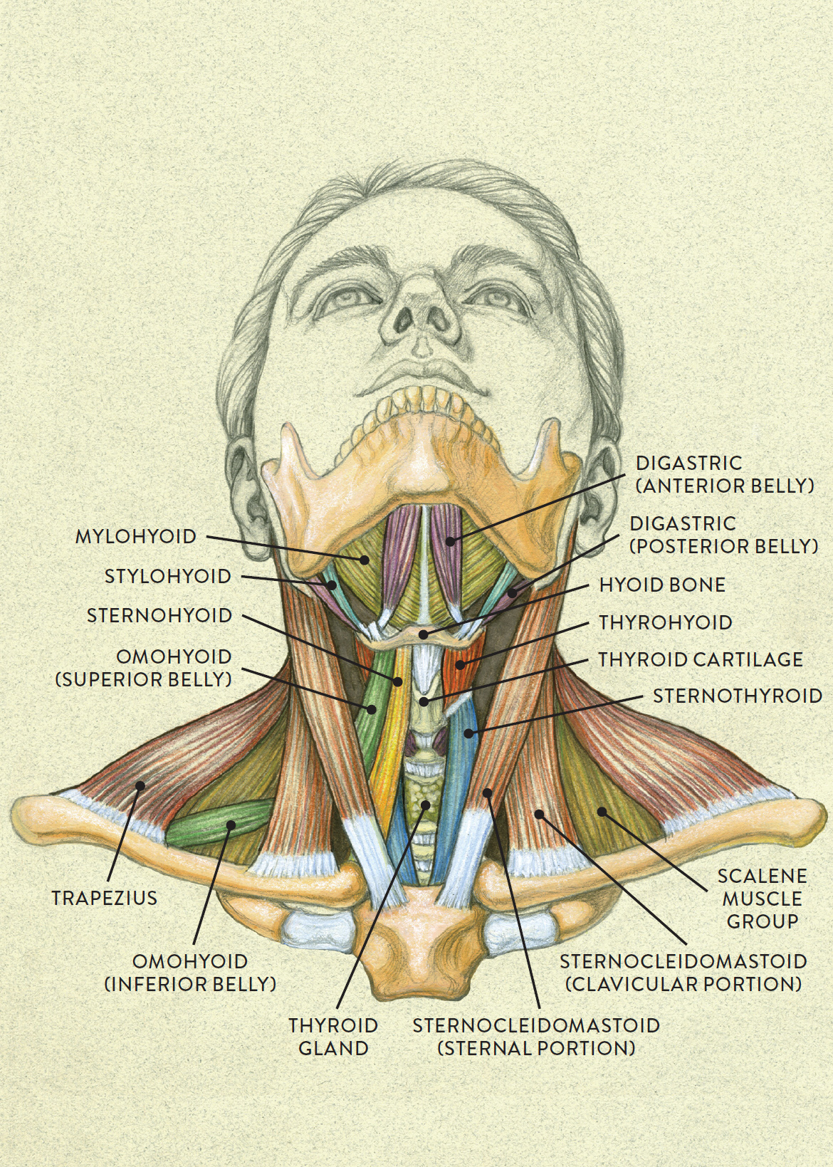
Anterior view of head tilting back from schoolbag.info The shoulder joint (glenohumeral joint) is a ball and socket joint between the scapula and the the resting tone of these muscles act to compress the humeral head into the glenoid cavity. Each deltoid muscle has three heads, or distinct parts: The shoulder muscles bridge the transitions from the torso into the head/neck area and into the upper extremities of the arms and hands. Deltoid (posterior fibers), teres major, teres minor, latissimus dorsi, pectoralis major (sternocostal fibers). Muscles of the shoulder can be subdivided into a variety of groups depending on origin, topography, function or innervation. Superficial layer with deltoid, trapezius, pectoralis. Printable shoulder muscles diagrams to help you study the muscles structure in human's shoulder. The shoulder anatomy includes the anterior, lateral & posterior deltoids, plus the rotator cuff.
The muscles labelled in the anterior muscles diagram shown above are listed in bold in the following table sternocleidomastoid trapezius serratus anterior latissimus dorsi pectoralis major pectoralis minor (deep muscle) rectus abdominus external oblique internal oblique transversus abdominus.
Although three ligaments protect and surround the shoulder joint, most of its stability comes from the powerful muscles and tendons of the rotator cuff. The anterior, lateral and posterior deltoid heads. Tutorials on the shoulder muscles (e.g rotator cuff muscles: If deltoid is paralysed, rounded contour of the shoulder is lost and there is loss of power of abduction of arm from 15 to 90ο. Anterior superior iliac spine insertion: There are around 650 skeletal muscles within the typical human body. Now label the diagram in your workbook! Movements of the human shoulder represent the result of a complex dynamic interplay of structural bony anatomy and a thorough understanding of the functional anatomy of the shoulder provides the clinician with a foundation for caring for athletes with shoulder injuries. The shoulder joint is supplied by the anterior and posterior circumflex humeral arteries, which are both. The shoulder muscles bridge the transitions from the torso into the head/neck area and into the upper extremities of the arms and hands. Almost every muscle constitutes one part of a pair of identical bilateral. But they do so from different angles. Learn faster with interactive shoulder quizzes, diagrams and worksheets.
Muscles of the shoulder can be subdivided into a variety of groups depending on origin, topography, function or innervation shoulder muscles diagram. Sternum and superior six the pectoralis major muscle is the most important muscle for the adduction and anteversion of the.